
Stay Ahead of the Curve in Radiology AI.
RadAISlice is your weekly intelligence briefing on the most critical developments at the intersection of radiology and artificial intelligence. Stop searching. Start leading.Subscribe to join 11k+ radiology professionals tracking the future of imaging AI.
Featured In

Lastest Issues
The latest developments in Radiology & AI.
Reports & Market Maps
Explore our deeper briefings on radiology AI trends, market structure, and event-level intelligence.
SIIM 2026 AI Landscape
Analysis of SIIM 2026 sessions, themes, speaker mix, and abstract activity through the lens of imaging AI.

ECR 2026 AI Product Landscape
A launch-level view of ECR 2026 product announcements: AI share, product mix, and modality focus.

ECR 2026 AI Industry Landscape
A data-driven map of ECR 2026 exhibitors: AI share, vendor mix, and regional patterns.
Your Weekly Slice of Innovation
Each issue is precisely structured to give you exactly what you need. No fluff, just facts and forward-looking insights.
Recent Industry News
The Latest Research
FDA Approvals Database
Latest News & Trends
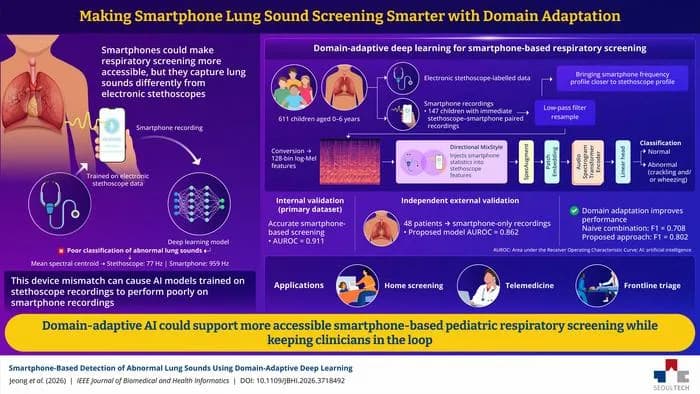
AI Framework Adapts Smartphone Audio for Pediatric Lung Sound Screening
Researchers developed a domain-adaptive deep learning model enabling smartphones to detect abnormal lung sounds in children for remote screening.

UC Irvine Implements AI to Find Lung Cancer Screening Candidates
UC Irvine's radiology department developed an NLP tool to identify patients eligible for lung cancer screening from EHR notes.

Webinar to Showcase Successful AI Automation in Radiology Workflows
A webinar will provide insights from imaging leaders on expanding and optimizing AI-driven automation in radiology clinical workflows.
From the Research Hub
AI-enhanced routine CT for opportunistic osteoporosis screening and risk assessment.
Osteoporosis-related fractures create a large and often preventable burden, yet many individuals undergoing CT for unrelated care do not receive skeletal assessment. This review evaluates artificial intelligence (AI)-enabled use of routine CT for opportunistic case-finding and risk assessment, with emphasis on the boundary between promising retrospective performance and safe clinical implementation. Automated vertebral segmentation and CT-derived bone-density estimation can reduce manual workload, and published diagnostic studies report high discrimination in selected cohorts. However, pooled estimates are heterogeneous because the reference standard, anatomical target, CT acquisition, reconstruction, contrast status, calibration method, prevalence, and validation design differ substantially across studies. These differences preclude a universal CT threshold or a universal minimum detector-row requirement; detector-row configuration should be considered together with the broader acquisition and reconstruction parameters. For implementation, AI should be deployed as a validated triage tool-not as a stand-alone diagnostic replacement for clinical assessment and indicated DXA/QCT. A credible pathway requires a defined CT-protocol eligibility check; pre-specified vertebral region of interest and exclusions; protocol- and contrast-aware quantification; scanner-specific quality assurance and calibration monitoring where applicable; external validation in representative populations; and an explicit route from an AI alert to confirmatory testing and treatment evaluation. AI may also enable concurrent vertebral-fracture and body-composition assessment, but clinical utility must be shown through downstream testing, treatment, fracture, workload, equity, and cost outcomes. The near-term priority is therefore not higher retrospective diagnostic performance alone, but reproducible, transparent, and prospectively evaluated integration into radiology workflows.
Real-World Validation of an AI-Based Opportunistic Osteoporosis Risk Stratification Tool Using Chest Radiographs in a Male-Predominant Cohort.
<b>Background:</b> Osteoporosis is a major public health concern and a leading cause of fragility fractures, yet screening with dual-energy X-ray absorptiometry (DXA) remains limited by accessibility and utilization. Opportunistic artificial intelligence (AI)-based analysis of routinely acquired chest radiographs (CXRs) may facilitate osteoporosis risk stratification. This study aimed to independently validate a regulatory-approved deep learning model (VeriOsteo™ OP) in a real-world, male-predominant cohort. <b>Methods:</b> We retrospectively analyzed 2336 patients who underwent both CXR and DXA at a medical center in 2023, including 1688 males (72.3%) and 648 females (27.7%). Osteoporosis was defined as a DXA T-score ≤ -2.5. AI performance was evaluated using sensitivity, specificity, positive predictive value (PPV), negative predictive value (NPV), accuracy, and area under the receiver operating characteristic curve (AUC), with sex-specific subgroup analyses. <b>Results:</b> The AI model achieved an accuracy of 76.97%, sensitivity of 69.67%, specificity of 84.64%, PPV of 82.66%, and NPV of 72.65%. The overall AUC was 0.854 (95% CI, 0.839-0.868). The AUC was 0.825 (95% CI, 0.794-0.854) in males and 0.849 (95% CI, 0.831-0.866) in females. PPV was lower in males than in females (60.00% vs. 83.39%), while other performance measures were broadly comparable. The optimal AI cutoff also differed between males (≤-1.9) and females (≤-2.5). <b>Conclusions:</b> The AI model demonstrated good discrimination for opportunistic osteoporosis risk stratification using CXRs in a real-world cohort. Its moderate sensitivity and lower PPV among males highlight potential sex-related differences in AI-assisted osteoporosis assessment. The model may be most appropriately used as an adjunctive risk-stratification and DXA-prioritization tool to identify individuals who may benefit from subsequent DXA evaluation rather than a standalone diagnostic test.
Multi-centre validation of MELD-graph for automated focal cortical dysplasia detection using 7T compared to paired 3T MRI
BackgroundSoftware tools can support MRI diagnosis of focal cortical dysplasias (FCDs) - subtle, surgically-treatable lesions that underlie drug-resistant focal epilepsy (DRFE). MELD-graph, a widely used open-source tool combining surface-based morphometry with a convolutional neural network, was developed using 1.5T and 3T MRI data. 7T MRI is increasingly used for lesion detection in DRFE. This study investigated whether MELD-graph is robust to domain shift at 7T. MethodsWe collated paired 3T and 7T T1-weighted MRI, and FLAIR where available, from 23 patients with FCD (6 histologically-verified, 17 identified radiologically) and 39 healthy controls at four epilepsy centres. Manual lesion labels were used as ground truth. MELD-graph was run on 3T images, on 7T images with the default pipeline, and on 7T images with adaptations for 7T MRI (SPM bias-field correction, SynthStrip skull-stripping, sub-millimetric FreeSurfer reconstruction), with and without ComBat harmonisation. FCD detection performance and surface morphometry features were compared. ResultsSensitivity for FCD detection was 65% (15/23) at 3T, 48% (11/23) for 7T default and 52% (12/23) for 7T adapted, with Combat harmonisation. Among radiologically 3T-negative patients, 2/8 were detected at 3T and 3/8 at 7T; and among histologically confirmed FCD, 3/6 at 3T and 1/6 at 7T. Specificity was significantly lower at 7T (default 39%, adapted 45%) than 3T (76%). False-positives predominantly occurred in the basal temporal lobes. ComBat harmonisation improved specificity in all conditions and largely compensated for field-strength dependent differences in surface morphometry features. Adding FLAIR at 7T increased false-positive predictions. ConclusionSensitivity is comparable but specificity of MELD-graph is lower at 7T compared to 3T. Adaptations for 7T MRI can reduce segmentation errors but do not significantly improve FCD detection, while harmonisation improves performance. The adapted pipeline is released open-source. 7T MELD-graph outputs may inform radiologist review in challenging cases but require cautious interpretation.
From the RadAI FDA Approvals Database
GE Medical Systems Ultrasound & Primary Care Diagnostics, LLC
Lung Guidance
Lung Guidance uses artificial intelligence to help clinicians optimize the process of capturing ultrasound images of the lungs, making it easier and more accurate to acquire diagnostic images for patient care.
Airs Medical, Inc.
SwiftSight-BodyComposition
SwiftSight-BodyComposition is a medical imaging software that analyzes MRI scans to assess body composition. This can help clinicians evaluate fat, muscle, and other tissue distributions, supporting diagnosis and treatment planning.
Brainlab SE
Spine Planning (3.0); Spine & Trauma Navigation; Alignment System Spine
The Brainlab Spine Planning (3.0), Spine & Trauma Navigation, and Alignment System Spine help clinicians plan and navigate spine surgeries using advanced image-guided systems. The software uses AI algorithms to automatically label vertebrae and assist in planning implant placement based on CT, MR, and X-ray images, making complex procedures more precise and efficient.
The Sharpest Insights, Effortlessly
Save Time
We scour dozens of sources so you don't have to. Get all the essential information in a 5-minute read.
Stay Informed
Never miss a critical update. Understand the trends shaping the future of your practice and research.
Gain an Edge
Be the first to know about the tools and technologies that matter, from clinical practice to academic research.
Ready to Sharpen Your Edge?
Subscribe to join 11k+ peers who rely on RadAISlice. Get the essential weekly briefing that empowers you to navigate the future of radiology.
We respect your privacy. Unsubscribe at any time.