
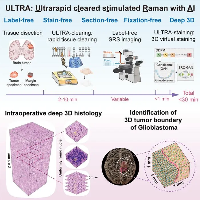
Researchers develop ULTRA, an AI-driven platform enabling 3D intraoperative tissue imaging for detecting glioma infiltration within 30 minutes.
Key Details
- 1ULTRA platform combines rapid tissue clearing, stimulated Raman scattering (SRS) microscopy, and AI-based virtual staining.
- 2Delivers 3D histology in approximately 30 minutes without conventional staining or sectioning.
- 3Evaluated using tissue from 17 glioma patients, covering various tumor subtypes.
- 4AI pipeline includes denoising, protein channel prediction, and H&E-like virtual staining modules.
- 5ULTRA outperformed 2D analysis, reducing 2D miss rates to 20%-23% and improving 3D CNN AUC to 0.965 vs. 0.909 for 2D.
- 6Currently at method development stage; not yet validated as a replacement for frozen sections or in large-scale clinical trials.
Why It Matters
This innovation addresses a key challenge in glioma surgery—rapid, accurate intraoperative tumor margin assessment—by providing rich 3D histological information in real time. The approach could accelerate intraoperative workflows and improve surgical precision if further validated clinically.

Source
EurekAlert
Related News

•EurekAlert
NIH Grant Funds AI Tool for Lung Cancer Discovery at Weill Cornell
Dr. Olivier Elemento receives an NIH Pioneer Award to develop an AI-human hybrid platform for cancer research, initially focusing on lung cancer using imaging and tissue data.

•EurekAlert
AI Model Combines ECG and Blood Tests to Spot Heart Transplant Rejection
An AI model integrating ECG data and blood biomarkers predicts heart transplant rejection with high specificity, reducing the need for invasive biopsies.
•EurekAlert
Study Shows AI Vision Misses Human-Like Perceptual Illusions
A York University study finds that AI vision systems lack the human-like perceptual shifts seen in biological vision during visual illusions.