
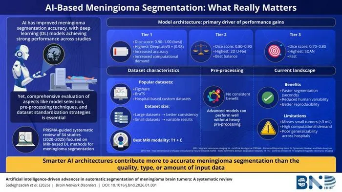
A systematic review highlights that AI model architecture, more than data or imaging specifics, drives improved tumor segmentation in brain MRI scans.
Key Details
- 1Researchers from the University of Auckland and affiliated institutes reviewed 34 studies on AI-based meningioma segmentation using MRI (2020-2025).
- 2Advanced AI architectures, such as DeepLabV3+, achieved the highest Dice scores (up to 0.98), indicating superior delineation performance.
- 3Model performance tiers were identified: Tier 1 (Dice 0.9-1, hybrid/advanced models), Tier 2 (Dice 0.8-0.9, e.g. 2D U-Net), Tier 3 (Dice <0.82, lightweight models).
- 4Data and imaging modality improvements contributed less to performance than advancements in AI architectures.
- 5Challenges remain, including underperformance for small tumors (<3 mL), high computational demands, and limited generalizability across hospitals.
- 6Most studies used contrast-enhanced T1 MRI; real-world deployment still faces workflow and infrastructure hurdles.
Why It Matters
This review provides critical evidence that prioritizing advanced AI architectures is key for accurate brain tumor segmentation in MRI, advancing radiology workflows and research focus. Understanding the limitations will help guide development of more deployable and generalizable models in clinical practice.

Source
EurekAlert
Related News

•EurekAlert
AI System Enhances Cancer Cell Detection via Light Scattering Spectra
Japanese researchers developed an AI system using light scattering spectra to improve cancer cell identification in cytology.

•EurekAlert
AI and X-ray Imaging Reveal Lost Texts in Ancient Roman Scrolls
AI and x-ray technology enable scientists to virtually read previously unreadable, carbonized Roman scrolls from Herculaneum.

•EurekAlert
AI’s Potential to Expand, Not Shrink, the Clinical Workforce
AI advancements may lead to more, not fewer, healthcare jobs, challenging common fears about workforce reductions in specialties like radiology.