
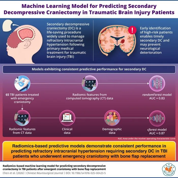
A radiomics-based machine learning model using CT scans predicts the need for secondary decompressive craniectomy after traumatic brain injury.
Key Details
- 1Researchers analyzed pre-evacuation CT scans from 65 adult TBI patients undergoing initial hematoma evacuation.
- 2Over 100 radiomic features were extracted and combined with demographic/clinical data for prediction.
- 3Radiomics-augmented models outperformed models using clinical data alone in forecasting the need for secondary decompressive craniectomy (DC).
- 4The multi-omic model showed improved predictive performance for refractory intracranial hypertension requiring further surgery.
- 5The study was led by Dr. Zhongyi Sun at Central South University and published in the Chinese Neurosurgical Journal.
Why It Matters
Radiomics and AI-enhanced CT imaging enable earlier identification of high-risk TBI patients, allowing more proactive neurosurgical care and potentially reducing complications. This approach highlights the growing integration of imaging AI in critical care and neurosurgery.

Source
EurekAlert
Related News

•EurekAlert
AI System Enhances Cancer Cell Detection via Light Scattering Spectra
Japanese researchers developed an AI system using light scattering spectra to improve cancer cell identification in cytology.

•EurekAlert
AI and X-ray Imaging Reveal Lost Texts in Ancient Roman Scrolls
AI and x-ray technology enable scientists to virtually read previously unreadable, carbonized Roman scrolls from Herculaneum.

•EurekAlert
AI’s Potential to Expand, Not Shrink, the Clinical Workforce
AI advancements may lead to more, not fewer, healthcare jobs, challenging common fears about workforce reductions in specialties like radiology.