
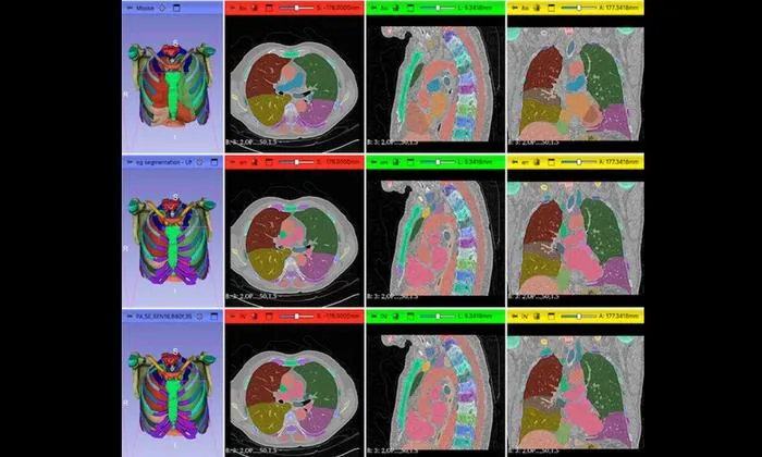
Researchers introduce an open-source approach for evaluating AI anatomy segmentation models in medical imaging without requiring ground truth annotations.
Key Details
- 1Six open-source AI segmentation models evaluated on chest CT scans from the National Lung Screening Trial (NLST).
- 2All model outputs standardized using DICOM format and harmonized medical lexicons (SNOMED-CV) for fair comparison.
- 3Strong agreement found across models for lung segmentation, but inconsistencies and systematic errors for bones and cardiac structures.
- 4Interactive visualization tools built on OHIF Viewer and 3D Slicer plugin enabled detailed side-by-side model review.
- 5Framework and datasets are open source; enables identification of reliable models and flags problematic cases.
Why It Matters
This framework addresses a major barrier for the radiology AI field by enabling fair, evidence-based model comparison in settings lacking expert-annotated ground truth. Such approaches help researchers select appropriate AI tools for large-scale studies and highlight the variability between widely used segmentation models.

Source
EurekAlert
Related News

•EurekAlert
AI System Enhances Cancer Cell Detection via Light Scattering Spectra
Japanese researchers developed an AI system using light scattering spectra to improve cancer cell identification in cytology.

•EurekAlert
AI and X-ray Imaging Reveal Lost Texts in Ancient Roman Scrolls
AI and x-ray technology enable scientists to virtually read previously unreadable, carbonized Roman scrolls from Herculaneum.

•EurekAlert
AI’s Potential to Expand, Not Shrink, the Clinical Workforce
AI advancements may lead to more, not fewer, healthcare jobs, challenging common fears about workforce reductions in specialties like radiology.