
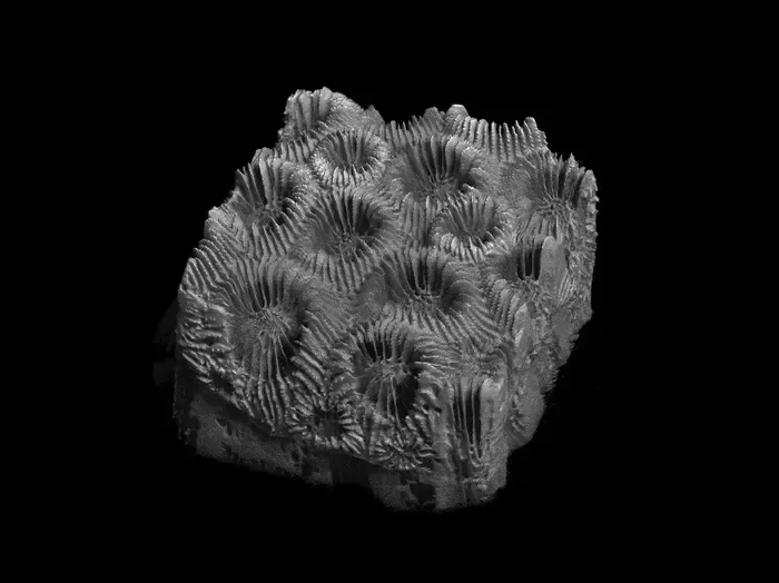
Researchers combined micro-CT imaging and deep learning to detect subtle disease-induced changes in coral skeletons with high accuracy.
Key Details
- 1Florida Atlantic University researchers used micro-CT imaging to generate detailed 3D reconstructions of coral skeletons.
- 2Deep learning, specifically U-Net family convolutional neural networks, was applied to segment and analyze skeletal features.
- 3The top-performing model (Attention U-Net) achieved more than 98% accuracy while reducing processing time to 7 hours.
- 4Study revealed disease-driven changes in porosity, density, and thickness of coral skeletons, providing new quantitative insights.
- 5The research opens the possibility of applying similar imaging-AI workflows to other biological, engineered, or geological samples.
Why It Matters
This work demonstrates the power of combining advanced 3D imaging with AI for high-throughput, non-destructive analysis of complex biological structures, a methodology that could be directly translated to radiology and imaging biomarker research. It also underscores how imaging AI can improve disease assessment and guide conservation or clinical strategies.

Source
EurekAlert
Related News

•EurekAlert
AI System Enhances Cancer Cell Detection via Light Scattering Spectra
Japanese researchers developed an AI system using light scattering spectra to improve cancer cell identification in cytology.

•EurekAlert
AI and X-ray Imaging Reveal Lost Texts in Ancient Roman Scrolls
AI and x-ray technology enable scientists to virtually read previously unreadable, carbonized Roman scrolls from Herculaneum.

•EurekAlert
AI’s Potential to Expand, Not Shrink, the Clinical Workforce
AI advancements may lead to more, not fewer, healthcare jobs, challenging common fears about workforce reductions in specialties like radiology.