
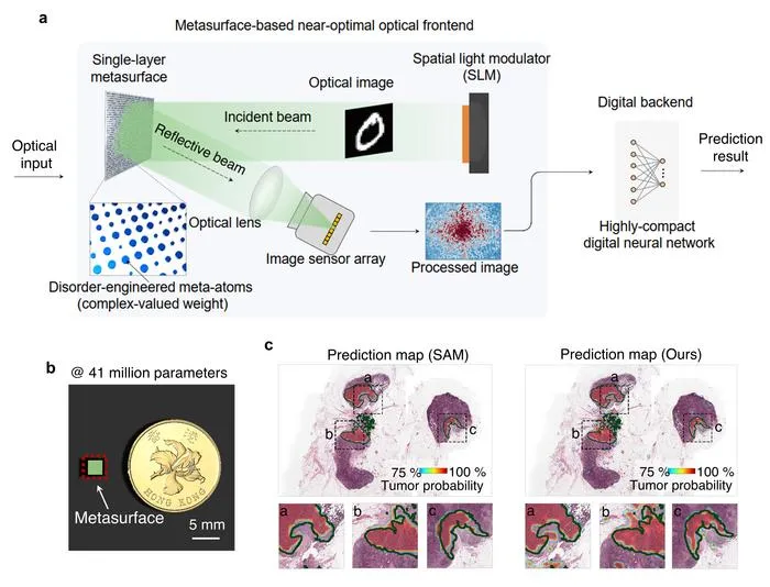
Researchers introduced a metasurface-based optical learning machine (MOLM) that achieves scalable, high-performance AI for imaging tasks including radiology and pathology.
Key Details
- 1The MOLM integrates 41 million optical parameters using a silicon metasurface, enabling ultra-wide optical neural networks.
- 2Performance matches state-of-the-art digital models on challenging computer vision tasks, including MNIST (99.2% accuracy), CIFAR-10 (91.6%), and medical imaging such as chest x-ray and cancer WSI analysis.
- 3In cancer whole-slide imaging, MOLM achieved results comparable to SAM (a top segmentation model), but in just 1.02 seconds versus 1.48 hours for SAM.
- 4The same device handled six different tasks (classification, segmentation, action recognition) without reconfiguration, showing broad versatility.
- 5System is highly robust to fabrication errors (±6% variation causing <1% accuracy drop), and works with incoherent illumination (over 98.3% accuracy across spectral bandwidths).
- 6In a clinical context, MOLM could process over 42,000 whole-slide pathology cases per day, versus only 8 for SAM, highlighting real-world impact.
Why It Matters
The MOLM’s drastic improvement in speed, robustness, and parameter scalability could fundamentally change how radiologists and pathologists use AI in practice, allowing analysis of massive datasets in real time with minimal hardware costs. This technology may enable wider adoption of AI in resource-limited healthcare settings and paves the way for next-generation imaging AI workflows.

Source
EurekAlert
Related News

•EurekAlert
AI System Enhances Cancer Cell Detection via Light Scattering Spectra
Japanese researchers developed an AI system using light scattering spectra to improve cancer cell identification in cytology.

•EurekAlert
AI and X-ray Imaging Reveal Lost Texts in Ancient Roman Scrolls
AI and x-ray technology enable scientists to virtually read previously unreadable, carbonized Roman scrolls from Herculaneum.

•EurekAlert
AI’s Potential to Expand, Not Shrink, the Clinical Workforce
AI advancements may lead to more, not fewer, healthcare jobs, challenging common fears about workforce reductions in specialties like radiology.