
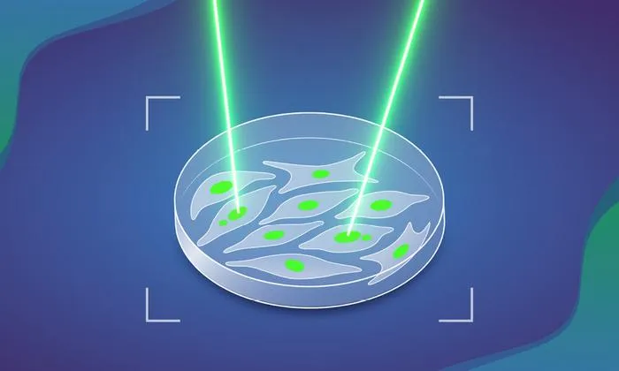
Researchers developed MAGIC, an AI-based system integrating automated microscopy and genomics to study chromosomal abnormalities linked to cancer.
Key Details
- 1MAGIC uses automated microscopy, AI-driven image analysis, and single-cell genome sequencing to identify and tag abnormal cells.
- 2The system can analyse nearly 100,000 cells in less than a day, vastly exceeding manual methods.
- 3Chromosomal abnormalities were seen in just over 10% of cell divisions, doubling with mutated p53 gene.
- 4MAGIC relies on a laser and photoconvertible dye to mark target cells, facilitating further analysis.
- 5The tool is versatile and can be trained to identify a variety of visually distinct cellular features.
Why It Matters
MAGIC represents a significant step forward in combining imaging AI and genomic methods for the high-throughput study of cellular abnormalities, offering potential advances in cancer detection and understanding cancer origins. Such AI-driven image analysis tools could pave the way for faster, more precise research and diagnostics in radiology and oncology.

Source
EurekAlert
Related News

•EurekAlert
AI System Enhances Cancer Cell Detection via Light Scattering Spectra
Japanese researchers developed an AI system using light scattering spectra to improve cancer cell identification in cytology.

•EurekAlert
AI and X-ray Imaging Reveal Lost Texts in Ancient Roman Scrolls
AI and x-ray technology enable scientists to virtually read previously unreadable, carbonized Roman scrolls from Herculaneum.

•EurekAlert
AI’s Potential to Expand, Not Shrink, the Clinical Workforce
AI advancements may lead to more, not fewer, healthcare jobs, challenging common fears about workforce reductions in specialties like radiology.