
Back to all news
Machine Learning and Fluorescence Microscopy Uncover Cellular Light Stress Responses
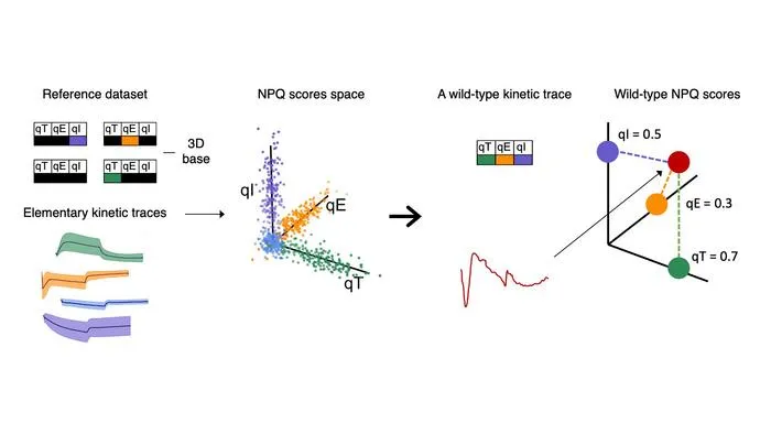
Researchers combined fluorescence microscopy and AI to analyze individual algae cells' responses to light stress, revealing previously hidden coordination in their protective mechanisms.
Key Details
- 1Custom automated fluorescence microscopy tracked hundreds of individual algal cells under light stress.
- 2Machine learning (dictionary learning, LDA) was used to distinguish three distinct NPQ (non-photochemical quenching) components at the single-cell level.
- 3Strong single-cell-level trade-offs were observed between protective mechanisms qE and qT, which bulk measurements cannot detect.
- 4Approach was non-destructive and adaptable to other photosynthetic organisms and stress types with detectable fluorescence signals.
- 5Integration with single-cell omics, flow cytometry, and microfluidics is suggested for broader biological and biotechnological application.
Why It Matters
This work demonstrates the potential of combining advanced imaging with AI for resolving cell heterogeneity—a concept highly relevant for precision medicine, radiology research, and the development of new imaging-based diagnostic tools.

Source
EurekAlert
Related News

•EurekAlert
AI System Enhances Cancer Cell Detection via Light Scattering Spectra
Japanese researchers developed an AI system using light scattering spectra to improve cancer cell identification in cytology.

•EurekAlert
AI and X-ray Imaging Reveal Lost Texts in Ancient Roman Scrolls
AI and x-ray technology enable scientists to virtually read previously unreadable, carbonized Roman scrolls from Herculaneum.

•EurekAlert
AI’s Potential to Expand, Not Shrink, the Clinical Workforce
AI advancements may lead to more, not fewer, healthcare jobs, challenging common fears about workforce reductions in specialties like radiology.