
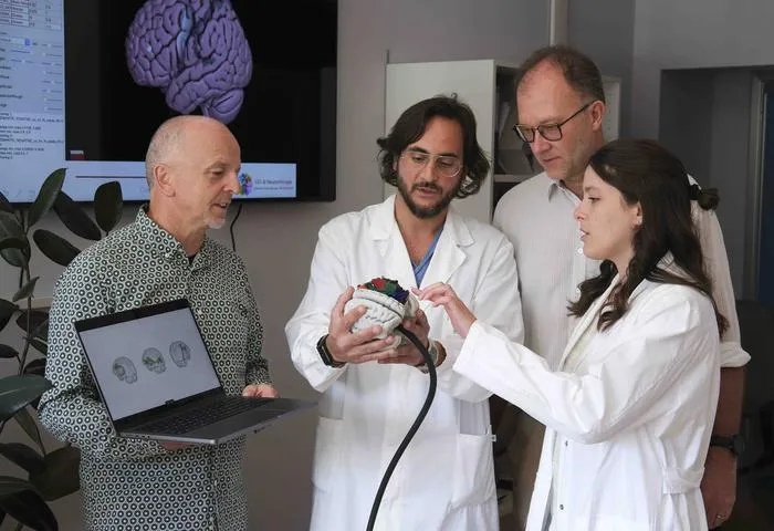
Researchers have developed BraDiPho, a tool that merges ex-vivo photogrammetric brain dissection data with in-vivo MRI tractography using AI.
Key Details
- 1BraDiPho enables precise, quantitative integration of ex-vivo brain dissections with in-vivo MRI scans and tractography.
- 2Thousands of high-resolution photographs are combined with AI to create detailed 3D models of white matter anatomy.
- 3The approach facilitates validation of tractography and personalized analysis of brain connectivity.
- 4Twelve anatomical specimens have been made freely available online for research and clinical use.
- 5BraDiPho supports neurosurgical planning, neuroscientific research, and teaching of brain anatomy.
Why It Matters
This innovation bridges a critical gap between anatomical validation and neuroimaging, enabling more accurate neurosurgical planning, improved interpretation of AI tractography, and widespread education. The freely available resource benefits radiology, neuroscience, and clinical AI communities by advancing methodology, precision, and transparency.

Source
EurekAlert
Related News
•EurekAlert
TRUECAM AI Framework Boosts Reliability in Cancer Pathology Imaging
PolyU researchers introduce the TRUECAM AI framework to enhance the trustworthiness of pathology AI for cancer diagnosis.

•EurekAlert
Deep Learning Shows Superior Sensitivity in Sinus Disease CT Changes
Automated deep learning achieves greater sensitivity in detecting treatment-related changes in chronic sinus disease on CT scans than standard visual scoring.
•EurekAlert
Mayo Clinic's AI Advances Ultrasound Detection of Heart Obstruction
Mayo Clinic researchers have developed an AI model that detects heart obstructions from routine ultrasound, potentially improving early identification of at-risk patients.