
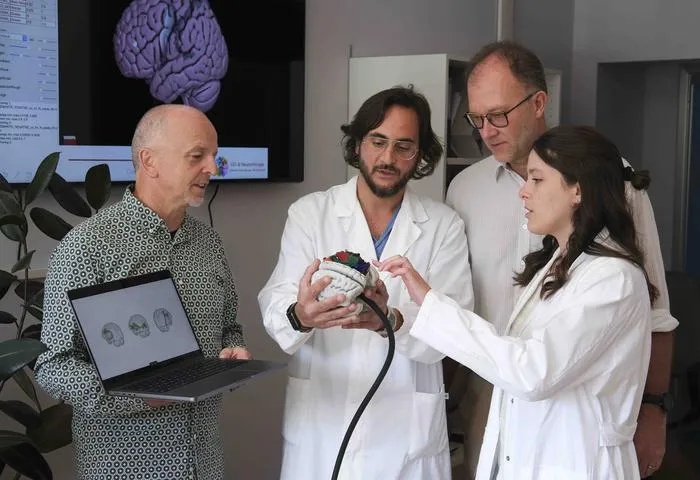
Researchers have developed BraDiPho, a tool that merges ex-vivo photogrammetric brain dissection data with in-vivo MRI tractography using AI.
Key Details
- 1BraDiPho enables precise, quantitative integration of ex-vivo brain dissections with in-vivo MRI scans and tractography.
- 2Thousands of high-resolution photographs are combined with AI to create detailed 3D models of white matter anatomy.
- 3The approach facilitates validation of tractography and personalized analysis of brain connectivity.
- 4Twelve anatomical specimens have been made freely available online for research and clinical use.
- 5BraDiPho supports neurosurgical planning, neuroscientific research, and teaching of brain anatomy.
Why It Matters
This innovation bridges a critical gap between anatomical validation and neuroimaging, enabling more accurate neurosurgical planning, improved interpretation of AI tractography, and widespread education. The freely available resource benefits radiology, neuroscience, and clinical AI communities by advancing methodology, precision, and transparency.

Source
EurekAlert
Related News

•EurekAlert
AI System Enhances Cancer Cell Detection via Light Scattering Spectra
Japanese researchers developed an AI system using light scattering spectra to improve cancer cell identification in cytology.

•EurekAlert
AI and X-ray Imaging Reveal Lost Texts in Ancient Roman Scrolls
AI and x-ray technology enable scientists to virtually read previously unreadable, carbonized Roman scrolls from Herculaneum.

•EurekAlert
AI’s Potential to Expand, Not Shrink, the Clinical Workforce
AI advancements may lead to more, not fewer, healthcare jobs, challenging common fears about workforce reductions in specialties like radiology.