
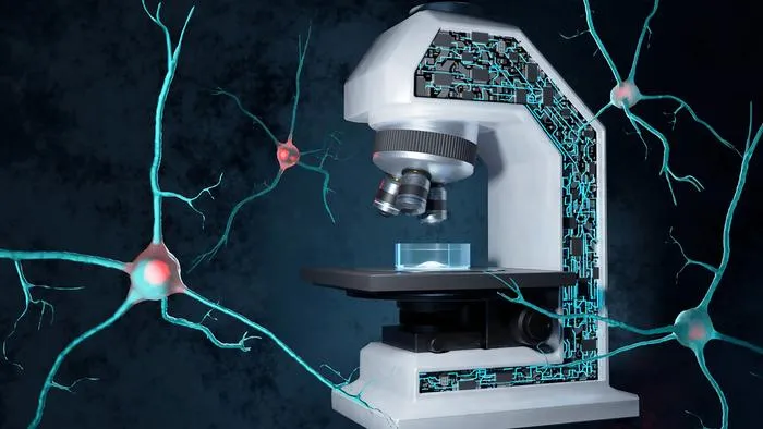
EPFL researchers created an AI-driven microscopy system that predicts and analyzes misfolded protein aggregation in real time.
Key Details
- 1Developed a self-driving imaging system combining multiple microscopy methods and deep learning.
- 2System predicts and detects protein aggregation—a hallmark of neurodegenerative diseases—in living cells.
- 3Uses label-free microscopy to minimize sample alteration and maximize imaging efficiency.
- 4Upon aggregation detection, system triggers Brillouin microscope to analyze biomechanical properties of aggregates.
- 5Aggregation onset detection achieved 91% accuracy using a specialized deep learning algorithm.
- 6Published in Nature Communications, with potential impact on drug discovery and precision medicine.
Why It Matters
This work showcases how AI-driven, label-free imaging can reveal dynamic, disease-relevant cellular processes in unprecedented detail, opening new pathways for diagnostic research and drug development in neurodegenerative diseases.

Source
EurekAlert
Related News

•EurekAlert
AI System Enhances Cancer Cell Detection via Light Scattering Spectra
Japanese researchers developed an AI system using light scattering spectra to improve cancer cell identification in cytology.

•EurekAlert
AI and X-ray Imaging Reveal Lost Texts in Ancient Roman Scrolls
AI and x-ray technology enable scientists to virtually read previously unreadable, carbonized Roman scrolls from Herculaneum.

•EurekAlert
AI’s Potential to Expand, Not Shrink, the Clinical Workforce
AI advancements may lead to more, not fewer, healthcare jobs, challenging common fears about workforce reductions in specialties like radiology.