
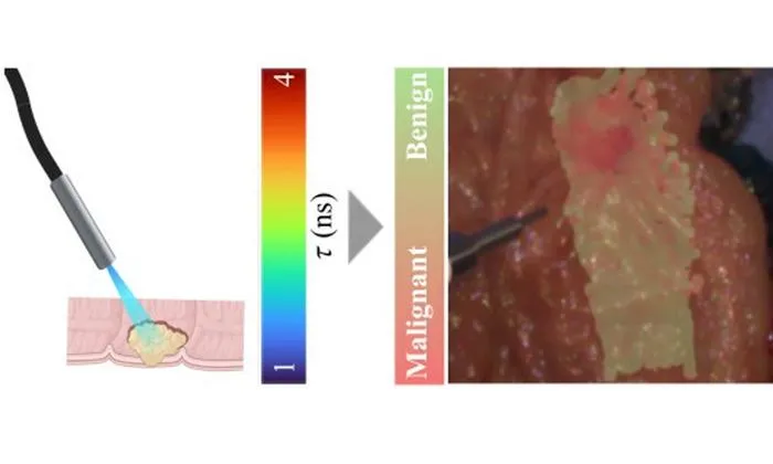
A label-free optical imaging technique using autofluorescence lifetime and AI can distinguish colorectal cancer with 85% accuracy.
Key Details
- 1Champalimaud Foundation researchers developed a fiber-optic, label-free optical imaging method for colorectal tissue analysis.
- 2Technique involves autofluorescence lifetime measurements at two wavelengths to capture biochemical differences.
- 3Machine learning (AdaBoost) trained on 117 patients' surgical specimens, validated with matched pathology results.
- 4On test data, the AI achieved 85% accuracy, 85% sensitivity, and 85% specificity.
- 5Potential applications include real-time cancer detection during colonoscopy or surgery, reducing the need for biopsies.
- 6Simplified versions of the imaging system delivered strong results, supporting future clinical use.
Why It Matters
Real-time, label-free optical imaging enhanced with AI could support faster, more accurate detection of cancer during endoscopic procedures, leading to earlier intervention and fewer unnecessary biopsies. This advances integration of functional imaging and AI in clinical workflows.

Source
EurekAlert
Related News

•EurekAlert
AI System Enhances Cancer Cell Detection via Light Scattering Spectra
Japanese researchers developed an AI system using light scattering spectra to improve cancer cell identification in cytology.

•EurekAlert
AI and X-ray Imaging Reveal Lost Texts in Ancient Roman Scrolls
AI and x-ray technology enable scientists to virtually read previously unreadable, carbonized Roman scrolls from Herculaneum.

•EurekAlert
AI’s Potential to Expand, Not Shrink, the Clinical Workforce
AI advancements may lead to more, not fewer, healthcare jobs, challenging common fears about workforce reductions in specialties like radiology.