
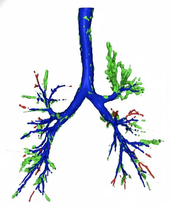
Southampton researchers developed an AI that surpassed radiologists in detecting hard-to-see airway obstructions on chest CT scans.
Key Details
- 1AI tool developed at University of Southampton detects radiolucent airway foreign bodies on chest CT.
- 2In study of 70 CT scans (14 positive for radiolucent FBA), radiologists detected 36% of cases; AI detected 71%.
- 3Radiologists had 100% precision but lower recall, while AI had 77% precision (some false positives).
- 4AI's F1 score (74%) exceeded that of radiologists (53%).
- 5Model tested on over 400 patient scans across three cohorts; aims to support, not replace, radiologists.
- 6Study published in npj Digital Medicine; research collaboration included UK and China.
Why It Matters
AI support can significantly improve detection of subtle, life-threatening foreign bodies in airways that are often missed on standard imaging, enhancing patient safety and outcomes. This work highlights the growing role of AI as an assistive tool in challenging radiological diagnoses.

Source
EurekAlert
Related News

•EurekAlert
AI Highlights Importance of Thymus Protection During Lung Cancer Radiotherapy
AI analysis reveals that radiation exposure to the thymus during lung cancer treatment may worsen patient outcomes.
•EurekAlert
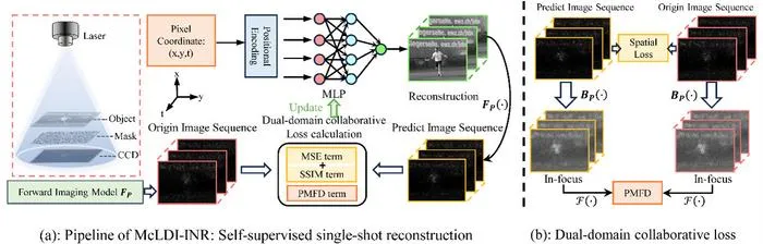
Deep Learning Enables Single-Shot High-Resolution Lensless Dynamic Imaging
Researchers from NJU and PKU have developed a lensless imaging method using deep learning that reconstructs high-fidelity dynamic images from single shots.

•EurekAlert
Weill Cornell Launches AI and Data Science Biomedicine Department Led by Dr. Dan Landau
Weill Cornell Medicine establishes a new Department of Systems and Computational Biomedicine, appointing Dr. Dan Landau as its first chair to drive AI-powered biomedical innovation.