
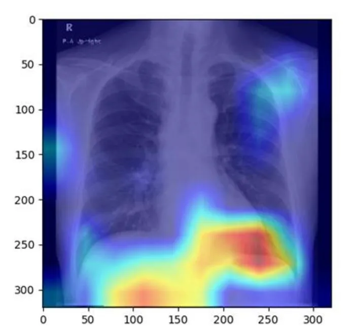
Researchers developed an AI model that can accurately detect fatty liver disease from routine chest X-rays.
Key Details
- 1The AI model was created by Osaka Metropolitan University researchers.
- 2It was trained and validated with 6,599 chest X-ray images from 4,414 patients.
- 3Model performance was strong, with AUC between 0.82 and 0.83.
- 4Chest X-rays are less costly and more commonly performed than ultrasounds, CTs, or MRIs currently used for liver diagnosis.
- 5Results were published in Radiology Cardiothoracic Imaging on June 20, 2025.
Why It Matters
This approach could enable earlier and more widespread detection of fatty liver disease using existing chest X-rays, reducing the need for more expensive or specialized imaging modalities. It demonstrates the potential for AI to repurpose common imaging studies for new clinical insights in radiology.

Source
EurekAlert
Related News

•EurekAlert
AI System Enhances Cancer Cell Detection via Light Scattering Spectra
Japanese researchers developed an AI system using light scattering spectra to improve cancer cell identification in cytology.

•EurekAlert
AI and X-ray Imaging Reveal Lost Texts in Ancient Roman Scrolls
AI and x-ray technology enable scientists to virtually read previously unreadable, carbonized Roman scrolls from Herculaneum.

•EurekAlert
AI’s Potential to Expand, Not Shrink, the Clinical Workforce
AI advancements may lead to more, not fewer, healthcare jobs, challenging common fears about workforce reductions in specialties like radiology.