
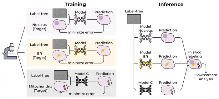
Researchers have developed a context-aware AI method to virtually label organelles in living cells using non-invasive microscopy.
Key Details
- 1Traditional fluorescence labeling damages living cells and has limitations for simultaneous staining.
- 2New AI uses not just image data, but also cellular context—such as cell shape, neighbors, and colony position—to improve labeling accuracy.
- 3The method enables accurate virtual staining of rare and dynamic processes like cell division, where previous approaches struggle.
- 4Published in Nature Methods (Dec 2025), developed at Ben-Gurion University of the Negev.
- 5The vision is a 'language model' for cells to generalize across cell types, microscopes, and conditions.
Why It Matters
This approach could revolutionize live-cell imaging by providing detailed and non-destructive visualization of cellular processes across various biological and clinical contexts. Improved virtual labeling enhances research and diagnostic capabilities in digital pathology, cell biology, and potentially clinical imaging.

Source
EurekAlert
Related News

•EurekAlert
AI System Enhances Cancer Cell Detection via Light Scattering Spectra
Japanese researchers developed an AI system using light scattering spectra to improve cancer cell identification in cytology.

•EurekAlert
AI and X-ray Imaging Reveal Lost Texts in Ancient Roman Scrolls
AI and x-ray technology enable scientists to virtually read previously unreadable, carbonized Roman scrolls from Herculaneum.

•EurekAlert
AI’s Potential to Expand, Not Shrink, the Clinical Workforce
AI advancements may lead to more, not fewer, healthcare jobs, challenging common fears about workforce reductions in specialties like radiology.