
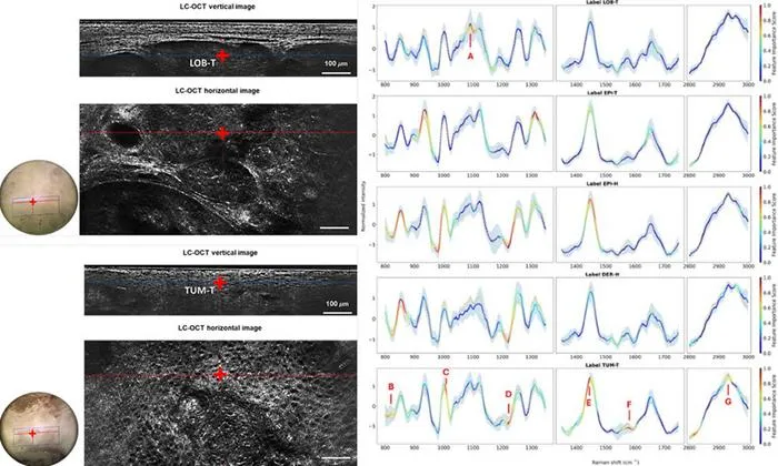
Researchers developed a dual-modality imaging system that combines high-resolution structural and chemical analysis with AI to improve skin cancer diagnosis.
Key Details
- 1The system merges line-field confocal optical coherence tomography (LC-OCT) with confocal Raman microspectroscopy.
- 2Over 330 nonmelanoma skin cancer samples were examined in a year-long clinical study.
- 3AI models trained on chemical spectra achieved classification accuracy of 0.95 for basal cell carcinoma and 0.92 for both basal and squamous cell carcinoma.
- 4The approach allows targeted, noninvasive analysis of suspicious skin structures at cellular and molecular levels.
Why It Matters
This innovation promises less invasive, more accurate skin cancer diagnosis by combining structural and biochemical imaging with AI classification, potentially reducing reliance on biopsies and improving patient outcomes.

Source
EurekAlert
Related News

•EurekAlert
AI System Enhances Cancer Cell Detection via Light Scattering Spectra
Japanese researchers developed an AI system using light scattering spectra to improve cancer cell identification in cytology.

•EurekAlert
AI and X-ray Imaging Reveal Lost Texts in Ancient Roman Scrolls
AI and x-ray technology enable scientists to virtually read previously unreadable, carbonized Roman scrolls from Herculaneum.

•EurekAlert
AI’s Potential to Expand, Not Shrink, the Clinical Workforce
AI advancements may lead to more, not fewer, healthcare jobs, challenging common fears about workforce reductions in specialties like radiology.