
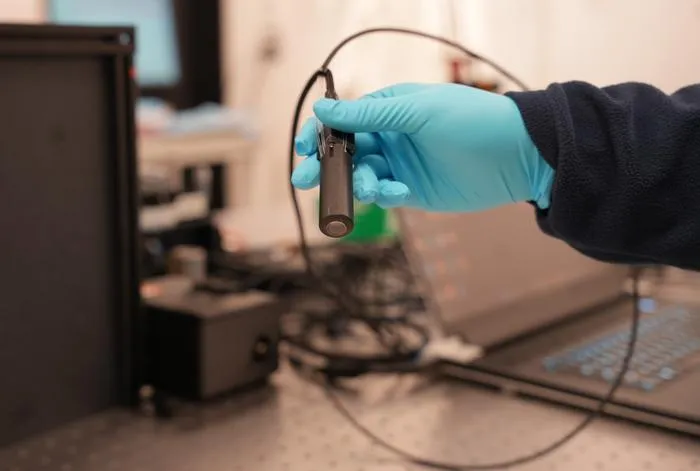
Researchers develop PrecisionView, a handheld AI-powered endomicroscope for real-time, high-resolution cancer diagnostics.
Key Details
- 1PrecisionView combines advanced optics and deep learning to image cellular and vascular structures non-invasively.
- 2Delivers a field of view five times, and depth of field eight times, greater than traditional in vivo microscopy.
- 3Displays real-time images at up to 15 frames per second, enabling clinicians to examine several square centimeters in one scan.
- 4Validated in healthy volunteers and human tissue samples, successfully identifying precancerous and cancerous lesions.
- 5The device is both compact (about the size of a pen) and low-cost (~$3,000), making it suitable for low-resource settings.
- 6Backed by Rice University, MD Anderson Cancer Center, NIH, NCI, and DARPA funding; larger clinical studies pending.
Why It Matters
This technology could significantly improve early cancer detection by making high-resolution microscopy portable, affordable, and usable at the point of care, especially in underserved regions. Integration of AI and hardware design sets a new paradigm for imaging devices in clinical oncology.

Source
EurekAlert
Related News

•EurekAlert
AI System Enhances Cancer Cell Detection via Light Scattering Spectra
Japanese researchers developed an AI system using light scattering spectra to improve cancer cell identification in cytology.

•EurekAlert
AI and X-ray Imaging Reveal Lost Texts in Ancient Roman Scrolls
AI and x-ray technology enable scientists to virtually read previously unreadable, carbonized Roman scrolls from Herculaneum.

•EurekAlert
AI’s Potential to Expand, Not Shrink, the Clinical Workforce
AI advancements may lead to more, not fewer, healthcare jobs, challenging common fears about workforce reductions in specialties like radiology.