

Researchers developed an AI model that uses routine CT scans to estimate a person's cardiovascular age.
Key Details
- 1Emory University's Empathetic AI for Heart Institute led the study.
- 2The AI focused on evaluating fat surrounding the aorta in chest CT exams.
- 3Over 48,000 chest CT examinations were analyzed; 4,000 for training, 44,000 for validation.
- 4Findings published in the European Heart Journal.
- 5The AI estimated individuals' 'true cardiovascular age' based on image analysis.
Why It Matters
This research demonstrates the power of AI in extracting novel, clinically relevant biomarkers from standard radiology scans. It could enable more personalized cardiovascular risk assessment without additional imaging, benefiting both diagnostic radiologists and preventive medicine.

Source
Cardiovascular Business
Related News
•Radiology Business
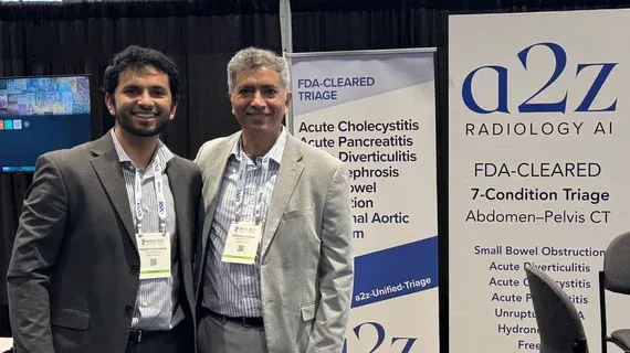
a2z Radiology AI’s Abdo-Triage Cleared for Expanded CT Findings
FDA has cleared a2z Radiology AI's Abdo-Triage tool to triage 10 additional findings on abdomen-pelvis CT scans.

•Radiology Business
AI Model Reduces Need for Gadolinium Contrast in Brain Tumor MRI
UCL researchers develop an AI tool to predict brain tumor MRI enhancement, reducing the need for gadolinium contrast agents.

•Radiology Business
Rad Partners Wins $1.29M FDA Grant for AI Radiology Report Evaluation Study
Cognita Imaging, part of Rad Partners, has received a $1.29M FDA grant to develop a new framework using large language models (LLMs) to evaluate AI-generated radiology reports.