
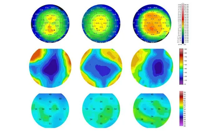
AI combined with polarization-sensitive OCT enables earlier and more accurate detection of subclinical keratoconus compared to standard tomography.
Key Details
- 1Researchers analyzed imaging data from 359 eyes using PS-OCT and compared it with two widespread corneal tomography devices (Pentacam, MS-39).
- 2AI models built on PS-OCT data were more effective in classifying subclinical keratoconus than shape-based tomography models.
- 3PS-OCT focuses on corneal collagen organization and sublayer thickness, revealing microstructural changes not captured by conventional imaging.
- 4Healthy and established keratoconus cases were classified similarly by all models; the main difference appeared for subclinical eyes.
- 5The research suggests PS-OCT provides complementary information to current imaging, improving the confidence of early keratoconus diagnosis.
Why It Matters
Detecting keratoconus at its subclinical stage can prevent progression and support safer refractive surgery decisions. Integrating advanced high-resolution imaging with AI may transform early eye disease diagnosis, reduce false positives in at-risk patients, and inform more precise clinical management.

Source
EurekAlert
Related News

•EurekAlert
AI System Enhances Cancer Cell Detection via Light Scattering Spectra
Japanese researchers developed an AI system using light scattering spectra to improve cancer cell identification in cytology.

•EurekAlert
AI and X-ray Imaging Reveal Lost Texts in Ancient Roman Scrolls
AI and x-ray technology enable scientists to virtually read previously unreadable, carbonized Roman scrolls from Herculaneum.

•EurekAlert
AI’s Potential to Expand, Not Shrink, the Clinical Workforce
AI advancements may lead to more, not fewer, healthcare jobs, challenging common fears about workforce reductions in specialties like radiology.