
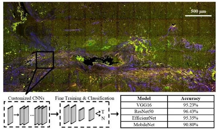
University of Arizona researchers combined label-free multiphoton microscopy with neural networks to accurately classify pancreatic neuroendocrine neoplasms in tissue samples.
Key Details
- 1Multiphoton microscopy (MPM) was used to image pancreatic neuroendocrine neoplasm (PNEN) samples without labeling.
- 2Researchers trained both traditional machine learning and four convolutional neural networks (CNNs) on these images.
- 3CNNs achieved classification accuracies ranging from 90.8% to 96.4%, outperforming the ML algorithm’s 80.6%.
- 4Analysis showed key features included collagen content and image texture metrics.
- 5The approach is faster than traditional pathology and was validated across samples from multiple biorepositories.
- 6Publication: Biophotonics Discovery, October 2, 2025, DOI: 10.1117/1.BIOS.2.4.045001.
Why It Matters
This study demonstrates the potential of advanced imaging and AI for real-time, highly accurate tumor detection, which could transform intraoperative pathology and improve surgical outcomes in oncology. Faster, label-free diagnosis may reduce delays and error in tumor identification during surgery.

Source
EurekAlert
Related News

•EurekAlert
AI System Enhances Cancer Cell Detection via Light Scattering Spectra
Japanese researchers developed an AI system using light scattering spectra to improve cancer cell identification in cytology.

•EurekAlert
AI and X-ray Imaging Reveal Lost Texts in Ancient Roman Scrolls
AI and x-ray technology enable scientists to virtually read previously unreadable, carbonized Roman scrolls from Herculaneum.

•EurekAlert
AI’s Potential to Expand, Not Shrink, the Clinical Workforce
AI advancements may lead to more, not fewer, healthcare jobs, challenging common fears about workforce reductions in specialties like radiology.