
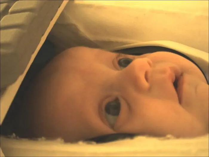
Neuroscientists used fMRI and AI models to show that infants as young as two months can categorise objects in their brains.
Key Details
- 1Study involved 130 two-month-old infants undergoing awake fMRI scans while viewing visual stimuli.
- 2Functional MRI tracked brain responses to 12 visual categories, including animals and objects.
- 3AI models were used to analyze and compare infant brain activation patterns for visual recognition.
- 4Research opens new opportunities for early neuroimaging and computational diagnosis in infants.
- 5Findings published in Nature Neuroscience highlight the foundation of visual cognition in early infancy.
Why It Matters
This is the largest awake fMRI study in infants to date and demonstrates the potential for imaging AI to investigate brain function and cognitive development at very early ages, with future implications for both clinical diagnostics and improving AI models. The cross-over between neuroimaging and computational models showcases the growing synergy in radiology and AI research.

Source
EurekAlert
Related News

•EurekAlert
AI System Enhances Cancer Cell Detection via Light Scattering Spectra
Japanese researchers developed an AI system using light scattering spectra to improve cancer cell identification in cytology.

•EurekAlert
AI and X-ray Imaging Reveal Lost Texts in Ancient Roman Scrolls
AI and x-ray technology enable scientists to virtually read previously unreadable, carbonized Roman scrolls from Herculaneum.

•EurekAlert
AI’s Potential to Expand, Not Shrink, the Clinical Workforce
AI advancements may lead to more, not fewer, healthcare jobs, challenging common fears about workforce reductions in specialties like radiology.