
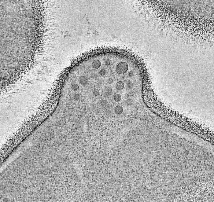
Researchers have discovered the ExHOS nanomachine responsible for constitutive exocytosis using advanced microscopy and AI-enhanced image analysis.
Key Details
- 1International team led by Pompeu Fabra University identifies ExHOS, the nanomachine controlling constitutive exocytosis.
- 2Discovery relied on integrating multiple advanced light and electron microscopes with artificial intelligence for image analysis.
- 3ExHOS organizes seven protein assemblies into a flexible ring, mediating molecular package delivery at the cell surface.
- 4ExHOS understanding has implications for infections, rare diseases, and cancer metastasis.
- 5Microscopy and AI enabled visualization of dynamic cellular machinery previously inaccessible by traditional imaging.
Why It Matters
The use of cutting-edge imaging and AI demonstrates the evolving power of bioimaging technologies, paving the way for deeper insights into fundamental biological processes and their medical implications. This integration is directly relevant to radiology and imaging AI professionals interested in high-resolution cellular and molecular imaging advancements.

Source
EurekAlert
Related News

•EurekAlert
AI System Enhances Cancer Cell Detection via Light Scattering Spectra
Japanese researchers developed an AI system using light scattering spectra to improve cancer cell identification in cytology.

•EurekAlert
AI and X-ray Imaging Reveal Lost Texts in Ancient Roman Scrolls
AI and x-ray technology enable scientists to virtually read previously unreadable, carbonized Roman scrolls from Herculaneum.

•EurekAlert
AI’s Potential to Expand, Not Shrink, the Clinical Workforce
AI advancements may lead to more, not fewer, healthcare jobs, challenging common fears about workforce reductions in specialties like radiology.