
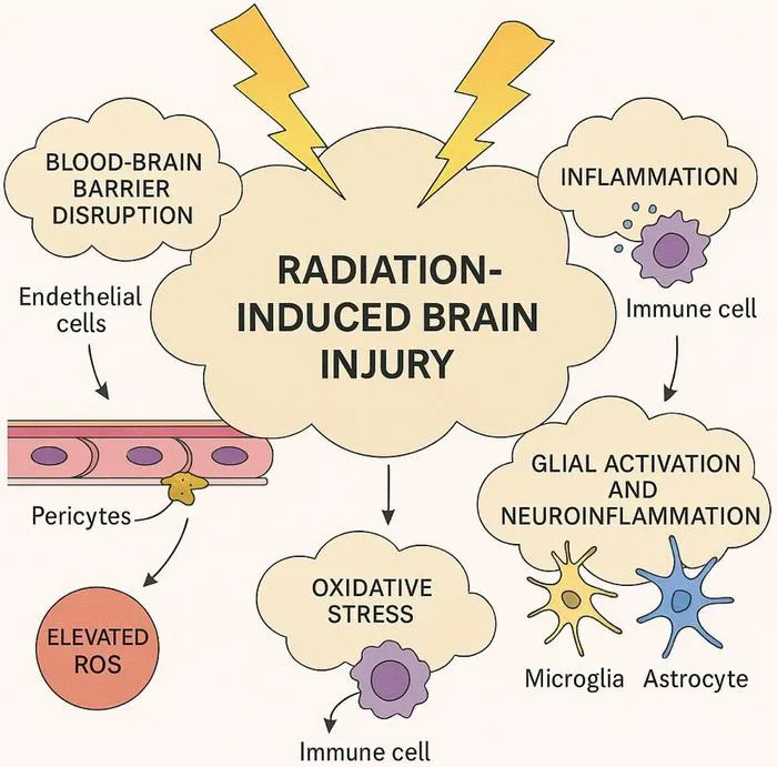
A state-of-the-art review highlights the use of multimodal imaging and AI to improve diagnosis and management of radiation-induced brain injury (RIBI).
Key Details
- 1Radiation-induced brain injury (RIBI) is a complex complication following cranial radiotherapy, impacting neurological function and quality of life.
- 2Multimodal imaging methods—including structural/functional MRI, diffusion and perfusion imaging, PET/CT, and radiomics—enhance early detection and differential diagnosis of RIBI versus tumor recurrence.
- 3AI techniques and machine learning models enable extraction of quantitative features, promising improved non-invasive diagnosis accuracy.
- 4Current interventions are shifting towards targeted, mechanism-driven therapies; Bevacizumab remains the only validated drug for radiation necrosis, while experimental approaches like stem cell therapy and neuromodulation are under study.
- 5Major challenges include lack of unified diagnostic criteria, early biomarkers, and seamless clinical integration of multimodal imaging and AI.
- 6The article calls for standardized protocols, expanded research, and multidisciplinary collaboration to achieve precision management.
Why It Matters
RIBI is an increasingly recognized complication given longer brain tumor survival, yet its diagnosis remains difficult using conventional imaging. Integrating advanced multimodal techniques and AI-driven analysis could transform early detection and precision treatment, representing a critical area for radiology and imaging AI innovation.

Source
EurekAlert
Related News

•EurekAlert
AI System Enhances Cancer Cell Detection via Light Scattering Spectra
Japanese researchers developed an AI system using light scattering spectra to improve cancer cell identification in cytology.

•EurekAlert
AI and X-ray Imaging Reveal Lost Texts in Ancient Roman Scrolls
AI and x-ray technology enable scientists to virtually read previously unreadable, carbonized Roman scrolls from Herculaneum.

•EurekAlert
AI’s Potential to Expand, Not Shrink, the Clinical Workforce
AI advancements may lead to more, not fewer, healthcare jobs, challenging common fears about workforce reductions in specialties like radiology.