

MD Anderson researchers developed an AI model that predicts which lung cancer patients may develop serious immunotherapy-induced pneumonitis using routine CT scans.
Key Details
- 1The AI model, called CIPHER, analyzed over 590,000 CT slices from 2,500 lung cancer patients.
- 2CIPHER predicts risk of checkpoint-inhibitor pneumonitis prior to treatment with an AUC of ~0.83 in both internal and external validation cohorts.
- 3The model outperformed conventional clinical risk models and radiomics approaches.
- 4It maintained accuracy across different patient populations, CT scanners, and protocols.
- 5Patients identified as high risk by the model developed pneumonitis earlier, indicating detection of underlying vulnerability.
- 6Study published in Journal for ImmunoTherapy of Cancer and funded by NIH, CPRIT, and institutional grants.
Why It Matters
Routine imaging can provide actionable insights on therapy-induced complications, aiding in personalized monitoring and intervention. Accurate pre-treatment risk stratification for pneumonitis could reduce patient morbidity and improve outcomes in lung cancer immunotherapy.

Source
EurekAlert
Related News
•EurekAlert
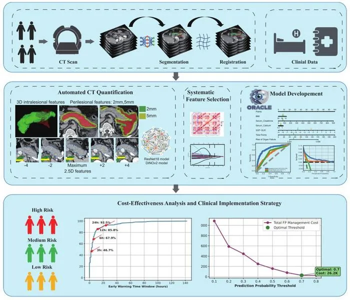
AI Model Predicts Organ Failure from CT in Pancreatitis Hours in Advance
A multicenter deep learning model (ORACLE) predicts organ failure in acute pancreatitis from CT scans with high accuracy, outperforming existing clinical tools.
•EurekAlert
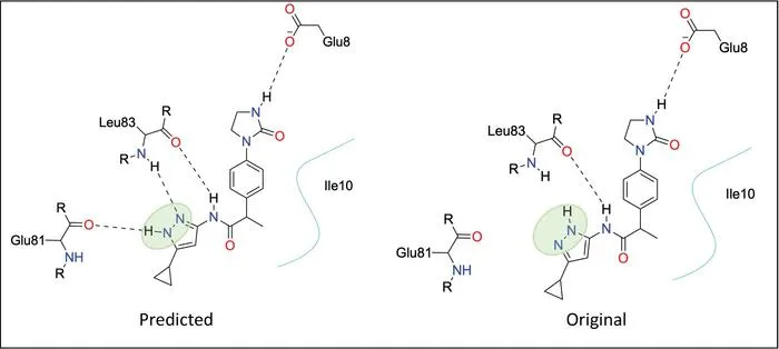
NYU Researchers Launch AI Tool for Stable Molecule Tautomer Prediction
NYU scientists have created an AI tool using crystallographic data to accurately predict stable tautomers in drug-like molecules.

•EurekAlert
Binghamton Researchers Secure AI Funding for Cancer Imaging Technologies
Binghamton University researchers earned SUNY funds to develop AI-powered cancer diagnostics using standard pathology imaging.