
A new AI framework (MENISA) combines CT scans, digital pathology, and tumor microenvironment data to predict survival risk for esophageal squamous cell carcinoma.
Key Details
- 1MENISA integrates clinical data, CT images, digital pathology, and seven tumor microenvironment markers.
- 2Retrospective study included 450 surgical patients from two Chinese hospitals.
- 3The model achieved a concordance index of 0.778 for internal validation and 0.675 in external hospital data.
- 4MENISA outperformed single-modality models in differentiating patient survival outcomes.
- 5Imputation methods improved robustness when CT annotations varied between institutions.
- 6Authors emphasize the need for larger, prospective validation before clinical adoption.
Why It Matters
Integrating multimodal imaging and pathology captures more biological detail, potentially enabling personalized risk assessments in cancer care. Validating such AI frameworks could transform prognostic strategies in radiology and oncology, if confirmed in larger studies.

Source
EurekAlert
Related News
•EurekAlert
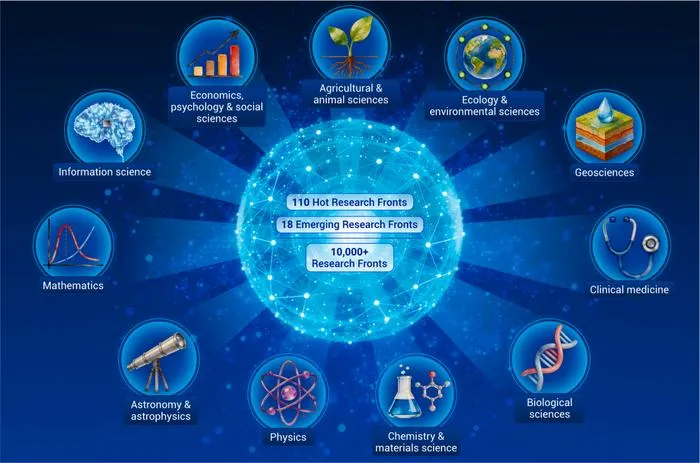
AI Now Drives Major Advances Across Scientific Fields, Report Finds
A comprehensive global analysis reveals AI as a cross-disciplinary engine, notably powering radiology and diagnostic innovations.
•EurekAlert
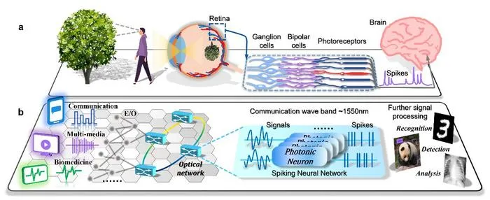
Photonic Encoder Transforms Medical Signals for Ultrafast AI Diagnosis
Researchers created a fiber-compatible, all-optical biomimetic spike encoder enabling rapid, precise conversion of medical signals for neuromorphic analysis.
•EurekAlert
Sydney Team Reveals Fractal Limits to Predictability in AI Training
University of Sydney researchers discover that deep learning models may remain unpredictable due to deep-rooted mathematical properties, even under controlled training conditions.